Lipid Fingerprints
BACLIB determines microbial species and antimicrobial resistance by measuring the lipids in microbial membranes. Each microbial species has a unique “fingerprint” pattern of membrane lipids. BACLIB detects a microbial species in a sample by detecting the unique lipid fingerprint of that species.
Evolutionary pressure for unique lipid fingerprints
Bacteria and fungi membranes are specialized for the organism’s ecological niche. Pathogens specialize their membranes to manipulate the host immune system and exploit the host. Importantly, pathogens gain an advantage by evading host immune responses that target a competitive pathogen. Consequently, pathogenic microbes exhibit unique lipid fingerprints, as has been demonstrated for a wide variety of microbes, including the most important human pathogens.
The membrane of Gram-negative bacteria is shown schematically below. Lipopolysaccharide (LPS) molecules are attached to the outer membrane. LPS consists of O antigen, core, and lipid A. O antigen is often highly variable, which aids bacteria in evading host detection. However, lipid A expression is unique to each Gram-negative species. BACLIB fingerprints for a Gram-negative bacterial species essentially comprise the lipid A expression for that species.
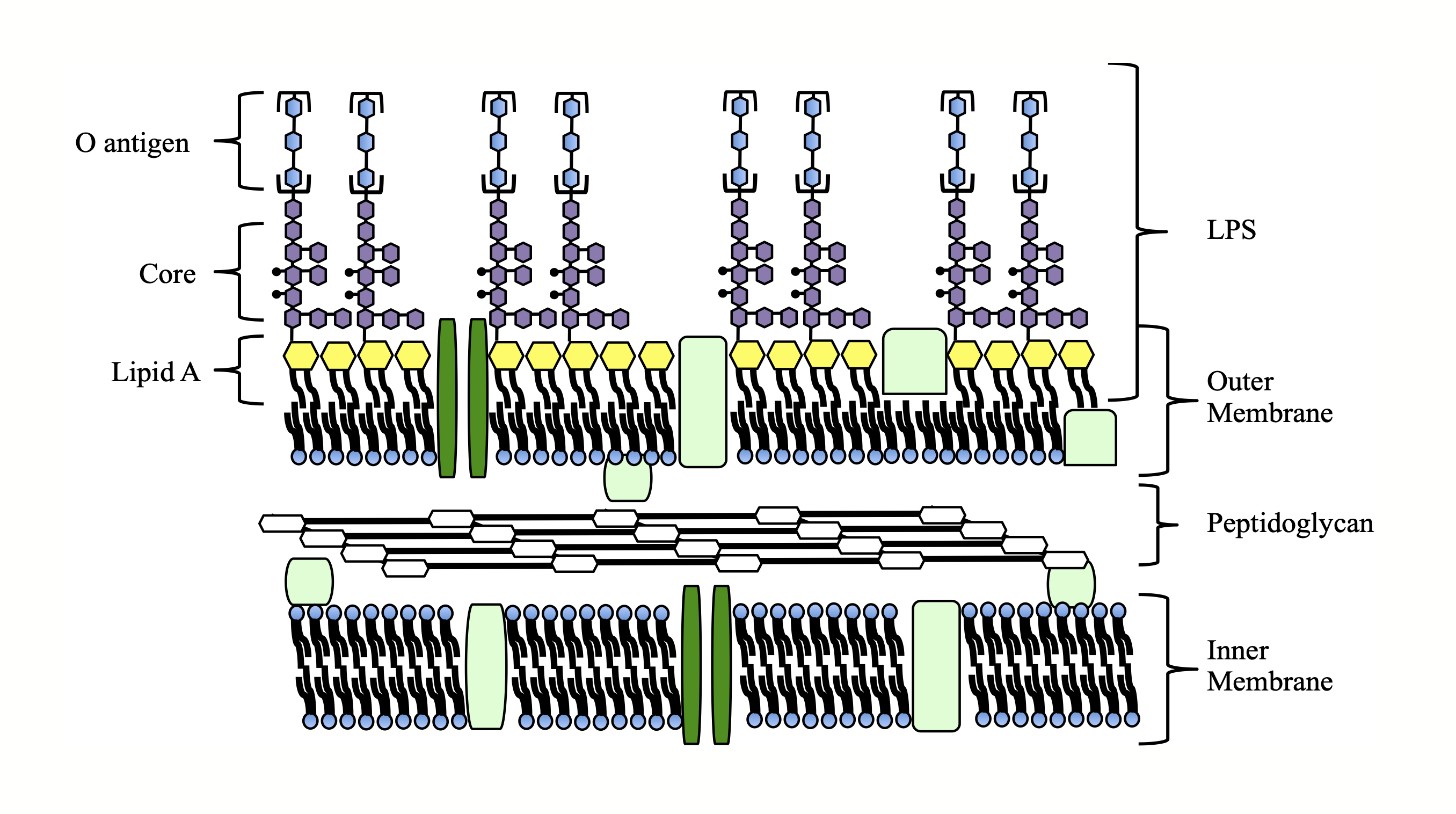
Gram-positive bacteria and fungi express different membrane lipids from Gram-negative bacteria, causing the BACLIB fingerprints of Gram-negative, Gram-positive, and fungal pathogens to be highly distinct. The same evolutionary pressures cause all three groups of pathogens to have BACLIB fingerprints that are unique per species.
Lipid A is species-specific
Over the last fifteen years, lipid A has been studied for a wide variety of Gram-negative bacteria. Each species exhibits a unique lipid A pattern. Individual species typically have a characteristic lipid A molecule that is unique to that species. Below are characteristic lipid A molecules for three common Gram-negative pathogens.
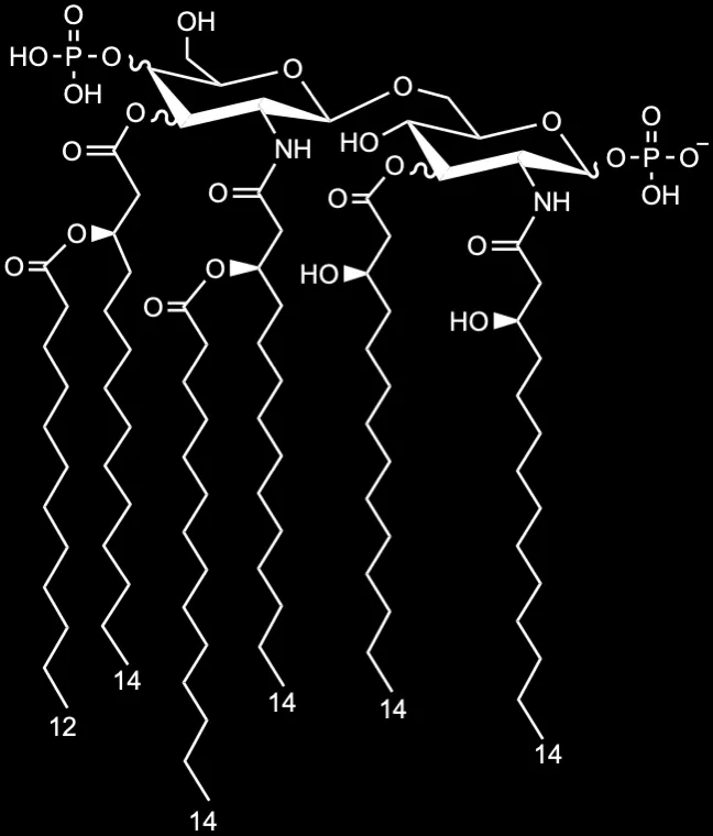
Escherichia coli
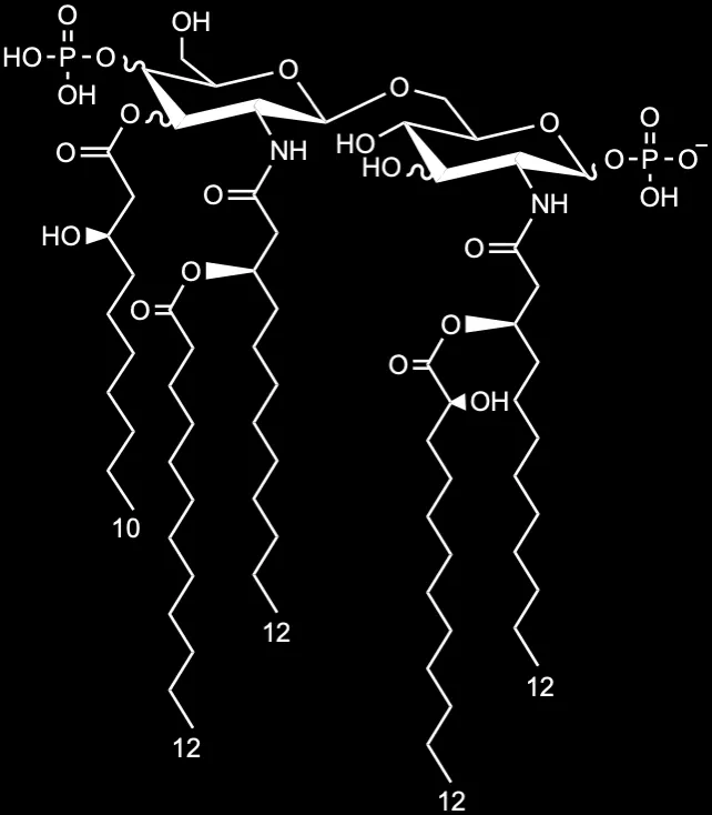
Pseudomonas aeruginosa
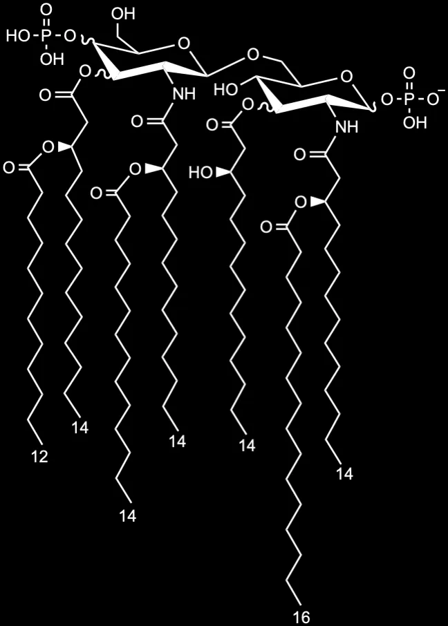
Salmonella typhimurium
Gram-positive bacteria have similar species-specific membrane patterns of lipoteichoic acid and cardiolipins. Fungi express species-specific sphingolipids and sterols in their membranes. Because BACLIB detects all of these membrane lipids, BACLIB identifies bacteria and fungi with one process.
Four species, four fingerprints
Escherichia coli
Gram-negative bacterium · lipid A
- Gram-negativelipid A
- Gram-positivecardiolipins and lipoteichoic acid
- Fungisphingolipids and sterols